HUMAN SKELETON
Introduction
The rigid parts of an animal that serve to support or protect the soft parts of the body constitute the
skeleton. There are two types of skeleton. (i) When the hard material is formed mainly on the outside of the body,
it is called exoskeleton. The muscles are attached to the inner surface of exoskeleton (e.g., insects, crabs, crustaceans
etc.). (ii) The endoskeleton is built within the body of the animals, surrounded by soft tissues, and have muscles
attached to their outer surface. It is made of bone and cartilage (e.g., vertebrate skeleton).
In vertebrates (e.g., man), the skeleton forms the internal framework of the body. The skeleton is important and
its basic functions are support, protection, movement and locomotion, muscle attachment, hemopoiesis (blood cell formation)
and storage of calcium and phosphate in bones.
Support:
The rigid skeleton supports the weight of the body, suspends some of the vital organs, and maintains
the shape of the body, despite vigorous muscular activity.
Protection: Certain delicate and important organs are protected against mechanical injury by a casing
of bones: for instance the brain, eyes, and inner ears are protected by the skull; the spinal cord by the vertebral
column ; and the heart and lungs by the thoracic cage.
Movement and locomotion: Many bones of the skeleton act as levers. When muscles pull on the levers
they produce movements, for instance the chewing action of the jaws, the breathing movements of the thoracic cage etc. Locomotion
is the result of coordinated action of muscles on the limb bones, to which they are attached by tendons.
Muscle attachment: To produce effective movement of any part of the skeleton, the muscles are attached
securely to it by flexible connective tissue bands called tendons or ligaments.
Hemopoiesis: The blood corpuscles are produced in the red bone marrow present in the spongy bones of
vertebrae and the sternum, scapula and in the ends of long bones, such as thehumerus and femur.
Storage of Calcium and Phosphate: which are released for several functions of the body.
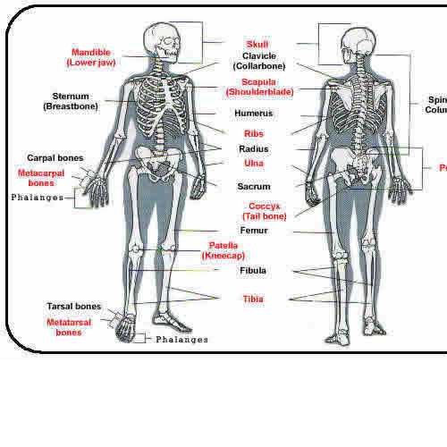
The adult human skeleton consists of 206 bones in the form of long bones (femur, humerus etc.);
short bones (wrist and ankle bones); flat bones (skull bones, ribs, shoulder blades) and irregular bones
(vertebrae, lower jaw, etc). The entire skeleton consists of two main parts: (A) the axial skeleton and (B) the appendicular
skeleton.
Axial Skeleton
The axial skeleton consists of the bones that form the upright portion or axis of the body. (i.e.
skull, ear bones, hyoid bone, vertebral column, sternum and ribs).
- The Skull: The skull is formed of 28 irregularly shaped bones (including inner ear bones). It consists of two
sets of bones cranial bones (brain case) and facial bones. Of the eleven paired and six single bones of the
skull, only the mandible (lower jaw bone) is movable, the other skull bones are joined together by immovable articulations:
called sutures.
At birth, many cranial bones are not fused (sutured) so that six spaces are left without any bony covering.
These spaces are called fontanels. These spaces allow change in the shape of the child’s head in passing through
the birth canal during birth, and allow for brain growth. By the second year of development, fontanels are completely fused.
The facial bones (14) consist of paired nasal, maxillae, zygomatic, palatine, lacrimal, inferior
nasal bones, and single mandible and vomer bones.
In adult males, the skull on average is larger, thicker and heavier, with larger air sinuses than
in adult females.
(B) Vertebral Column:
It forms the central axis of the body with the skull resting upon it. It consists of
33 irregular
bones called
vertebrae, joined to each other to
support the trunk; it allows a good deal of
movement,
provides
articulation with ribs and pelvic bones and
protects the spinal cord. The vertebral column is about
28 inches long and shows cervical, thoracic and lumbar
bends or
curvatures
All the vertebrae are constructed with the same basic structure and have two parts: (i) the body
or centrum (i.e. the anterior or ventral part) and ; (ii) the neural arch (i.e. the posterior or dorsal
part). The vertebral canal lies between the body and arch, and encloses the spinal cord. The arch bears three processes
for attachment of muscles; two transverse processes, one on either side that articulates with ribs, and a spinous
process which projects dorsally to which muscles and ligaments are attached. The arch also bears articular processes-(two
superior and two inferior) which project rostrally and caudaully respectively. These processes articulate with each other
on adjacent vertebrae. Between the successive vertebrae are a pair of small openings called intervertebral foramina for
the passage of spinal nerves. Between each of the vertebrae are cartilaginous pads; the intervertebral discs.
The vertebrae are named according to the region in which they lie (i) Cervical-7, (ii) Thoracic-12 (iii)
Lumbar-5 (iv) Sacral-5, fused into one sacrum and (v) Coccyl-4, fused into one: the Coccyx.
i) Cervical vertebrae:
Of the seven cervical vertebrae the third, fourth, fifth and sixth present similar
features, hence are called the
typical cervical, while the first, second and seventh present special features, hence
are called
atypical.