|
The Nervous System
NERVOUS CO-ORDINATION
Introduction
The behavior of an animal in its environment to maintain itself depends on the coordination of its organs
systems. Without coordination of various organ systems, various physiological processes would work in a haphazard way, without
linking together activities. The linking together in time and space of various activities of an animal is called coordination.
Coordination is brought about by the nervous system and sense organs, and by means of chemical substances (hormones) secreted
by the endocrine glands.
During the course of evolution, nervous systems have become more complex. This is largely because animals
have become larger and more mobile, requiring more neurons than a sedentary animal. The most fundamental function of a nervous
system is (1) to receive a stimulus (2) transmission of a stimulus to a central "brain", (3) interpretation and analysis of
the stimulus and (4) proper response by an effector.
With the evolution of a complex nervous system and bilateral
symmetry, cephalization (formation of a head) has taken place, and the ganglia in the head became large enough to be
called a brain, which is the main nervous control center of the body.
The Human Nervous System : The human nervous system is divided into two interrelated parts:
(1) The central nervous system (brain and spinal cord).
(2) The peripheral nervous system (nerves and ganglia).
Central Nervous System
(A) Brain
The human brain weighs about 1.5kg (average 1350 gms or 3 pounds) and is enclosed in the cranial (skull)
cavity. It is covered by 3 membranes, called meninges. The outer membrane, called dura mater ("tough mother")
is a tough, protective covering formed of white fibrous tissue the middle delicate membrane is called arachnoid (web
like), and the inner most transparent membrane is called pia mater ("soft mother"), containing blood vessels (nutritive
in function). Between the bony and membranous coverings, there is cerebrospinal fluid, which acts as a protective internal
cushion. The bony skull, meninges and the cerebrospinal fluid protect the brain from external injury and shocks.
- Forebrain
: It consist of two divisions, the anterior telencephalon with olfactory lobes, cerebral hemispheres and
the posterior diencephalon with thalamus and hypothalamus.
The cerebrum is the largest area of the brain and is divided by a longitudinal fissure into two cerebral
hemispheres. Each hemisphere consists of a frontal lobe, parital lobe, occipital lobe and temporal lobe. It shows prominent
fissures (deeper furrows) or sulci (shallow grooves) and gyri or convolutions (thin ridges between sulci). Structurally, the
hemispheres are made up of 3 portions (i) Cerebral cortex or grey matter, (ii) Cerebral tracts are bundles of
axons located inside the brain and extend in three directions forming projection fibers, association fibers, and transverse
fibers forming the corpus callosum (iii) Basal nuclei or ganglia located deep within the cerebral hemispheres,
connected to each other and with the thalamus and hypothlamus.
CEREBELLUM
b) The thalamus is a large, oval part located above the midbrain. All sensory impulses (except for
olfactory sensations) entering the cerebrum must pass through the thalamus.
c) The hypothalamus forms the floor and lower part of the brain formed by the thickening of the diencephalon. Part
of the hypothalamus also extends downwards and contributes to the pituitary body. Here, the nervous and hormonal systems interact.
It is concerned with different bodily activities, homeostasis, and is the center for the parasympathetic nervous system. Internally,
the downgrowth from the diencephalon forms the pituitary body, while the thin and vascular roof (anterior choroid plexus)
secretes cerebrospinal fluid into the third ventricle.
2) The midbrain is a short, constricted mass mostly of white matter surrounding a central cavity.
Ventrally, it connects the pons and cerebellum with the cerebral hemispheres by tracts of nerve fibers. In mammals, the analysis
of vision has moved out of the midbrain and has become a function of the forebrain. The dorsal part of the midbrain has four
small rounded projections which contain visual reflex (superior colliculi) and auditory reflex centers (inferior colliculi).
3) The hindbrain or rhombencephalon consists of two subdivisions, anterior metencephalon and
a posterior myelencephalon. The hindbrain has 3 main parts : (a) cerebellum (b) medulla and (c) pons.
(a) The cerebellum occupies the lower posterior part of the skull cavity. It is the second largest
part of the brain and consists of the outer layer of grey matter (cortex) and an inner core of white matter (medullary body).
Its surface is also grooved, but the convolutions are less prominent than cerebral hemispheres. The cerebellum has two large
lateral masses, the cerebellar hemispheres, and a central portion, the vermis. The internal white matter has
short and long nerve tracts, which connect various portions of the cerebellum with each other, and with other parts of the
brain and spinal cord.
(b)The medulla (medulla oblongata), also called the "brainstem" is the anterior enlargement
of the spinal cord, and consists of ascending and descending tracts of white matter. The vital nuclei in the medulla
control vital reflex centers such an heart action, respiration, diameter of blood vessels, etc. The non vital nuclei
contain reflex centers for activities such as vomiting, coughing, swallowing, sneezing, etc.
(c) The pons or pons varioli lies just above the medulla and is composed of white matter. It
contains transverse tracts that conduct impulses between the cerebellum, medulla and cerebrum. In pons, the nerve tracts between
brain and the spinal cord cross from the left side to the right side. Because of this crossover, the left side of the brain
controls activities of the right side of the body and vice versa.
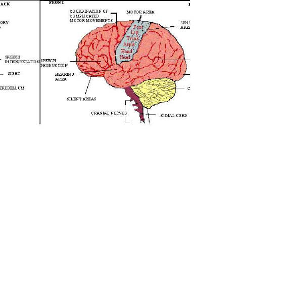
Functions of different parts of the brain
1) The cerebral hemispheres (cerebrum) perform (a) all mental functions, reason, will, memory and
intelligence, and (b) many essential motor sensory and visceral activities. Certain functional areas of cerebral cortex like
sensory, motor and association areas are mapped out after experimentation; these control motor, sensory and other activities
(Figure 23.3). The portion of cerebrum that governs muscular movements is known as the motor areas; those involved
in analysis of sensations are called sensory areas and those concerned with the higher faculties such as reasoning,
will, understanding, memory, personality, ethics, etc., are called association areas.
2) Basal ganglia or nuclei play a role in producing movement.
3) The thalamus is associated with pain, pleasure and emotions. It functions as an arousal or alert
mechanism of the body. It also has a role in producing complex reflex movements.
4) The hypothalamus is the control center for homeostasis, partial control of sleep and wakefulness,
regulation of body temperature and food intake, and controls of metabolism and water balance through synthesis of ADH secreted
by pituitary, regulation of autonomic activities, and control of various reproductive functions.
5) The pituitary body is the master endocrine gland.
6) The cerebellum coordinates the voluntary skeletal muscle activities and several other brain centers.
It controls skeletal muscles related to maintenance of equilibrium, smooth, timed, precise and steady body movements.
7) The medulla is the automatic control center for the heart beat, breathing, swallowing, sneezing,
etc. Sleep and loss of consciousness and cerebrum activities are controlled by tissues within the medulla.
8) The pons serves as a reflex center, regulates respiration, and serves as a conduction pathway between
the spinal cord and the brain.
Functional areas of the brain
B) The Spinal Cord
The spinal cord extends from the medulla or brain stem down to the second lumbar vertebra. It is surrounded
and protected by the meninges (same as covering the brain) and the vertebrae of vertebral column. It is a hollow, oval shaped
cylinder, tapering slightly as it descends.
Functions : (1) The spinal chord serves as a pathway for the condition of impulses between the receptors
and effector organs and the brain. (2) It acts as a reflex center (spinal reflexes) for many local simple reflexes.
(i) Cranial nerves : There are 12 pairs of cranial nerves in mammals (10 pairs in fishes and amphibians)
and most of them lead away from the medulla oblongata. Of these 12 pairs, some are purely sensory, some are purely motor and
the others are mixed. The table shows the name, number, nature, function and distribution of the cranial nerves.
Cranial nerves of man
|
No. |
Name |
Nature |
Function |
Distribution |
|
1) |
Olfactory |
Sensory |
Smell |
From nose |
|
2) |
Optic |
Sensory |
Vision |
From eye |
|
3) |
Oculomotor |
Motor |
Eye movement |
To muscles of eye ball |
|
4) |
Trochlear |
Motor |
Eye movement |
To muscles of eye ball |
|
5) |
Trigeminal |
Mixed |
Sensitive and jaw movement |
From and to face, teeth, lips, tongue,jaws |
|
6) |
Abducens |
Motor |
Eye movement |
To muscles of eye ball |
|
7) |
Facial |
Mixed |
Taste sensation, jaw movement |
From taste buds, to sali vary glands and face |
|
8) |
Auditory |
Sensory |
Hearing and balance |
From ear |
|
9) |
Glossopharyngeal |
Mixed |
Muscle movement and sensations |
From and to pharynx, from taste buds Salivary glands |
|
10) |
Vagus |
Mixed |
Sensory to chest and abodomen |
From and to visceral organs |
|
11) |
Spinal |
Motor |
Movement of shoulder muscles |
To shoulder muscles |
|
12) |
Hypoglossal |
Motor |
Movement of tongue |
To tongue |
ii) Spinal nerves : There are 8 cervical, 12 thoracic, 5 lumbar, 5 sacral and 1 coccygeal pairs of spinal nerves
(31 pairs in all), which leave the spinal cord between adjacent vertebrae. Each spinal nerve is formed by the joining of two
roots, a dorsal root (somatic and visceral sensory) and a ventral root (somatic and visceral motor), to form a mixed nerve
The Autonomic Nervous System
The central nervous system controls almost all voluntary activities of the animal. The autonomic nervous
system controls automatic activities which are free of will (involuntary such as heart beat, breathing, peristalsis
etc.) Thus, this system deals with the internal environment of the body (homeostasis). The autonomic nervous system is connected
with the central nervous system and is divided into two parts. (1) Sympathetic nervous system and (2) Parasympathetic
nervous system. The main structural and functional similarities and differences are given in the following table.
|
|
Sympathetic Nervous System |
|
|
1 |
Contains nerve fibers on which there is no voluntary control. (Involuntary nerve fibers.) |
1 |
---------- same ---------- |
|
2 |
Nerve fibers emerge from the brain or spinal cord. |
2 |
---------- same ---------- |
|
3 |
The final nerve fibers supplying organ do not have thick myelin sheath, thus sometimes called non-myelinated fiber. |
3 |
---------- same ---------- |
|
4 |
The ganglia lie close to the vertebrae/spinal cord. |
4 |
The ganglia are embedded in the wall of effector itself. |
|
5 |
Pre-ganglionic fibers are short. |
5 |
Pre-ganglionic fibers are long. |
|
6 |
Post-ganglionic fibers are long. |
6 |
Post-ganglionic fibers are short. |
|
7 |
Sympathetic fibers are generally activatory. (i.e. increase rate of heart beat.) |
7 |
Parasympathetic fibers are generally inhibitory (i.e. decreases rate of heart beat). |
|
8 |
Functions antagonistic to para-sympathetic fibers. |
8 |
Functions antagonistic to sympathetic fibers.. |
|
9 |
Terminals of post-ganglionic fibers secrete adrenaline or epinephrin (adrenergic fibers). |
9 |
Terminals of post ganglionic fibers release acteylcholine (cholinergic fibers.) |
|